Choosing the best Suture Anchor System is not a simple matter of selecting the newest device. The right choice depends on tissue quality, tear pattern, surgical technique, and the patient’s healing environment. A small anchor may suit a compact repair, while a larger construct may provide stronger fixation in demanding tissue. Details matter. Even insertion angle can influence pullout resistance.
Dr. James R. Andrews has said, “Every patient is different, and every operation must be individualized.” That principle remains highly relevant when comparing anchor materials, thread configuration, knotless designs, and deployment methods. A surgeon should also examine evidence from clinical studies, manufacturer instructions, training requirements, and long-term outcomes. Marketing language can sound convincing. It is not always enough.
The best Suture Anchor System should support secure fixation without creating unnecessary complexity. It should match the procedure, the anatomy, and the surgeon’s experience. Handling matters in the operating room. So does consistency. A device that performs well in testing may feel different in dense or fragile tissue. That difference deserves attention.
There is no universal winner.
This guide explores the practical questions behind a responsible selection. It considers strength, biocompatibility, insertion control, imaging compatibility, and cost. Some choices will remain imperfect. That is worth admitting. Careful judgment, not fashionable design, should guide the final decision.
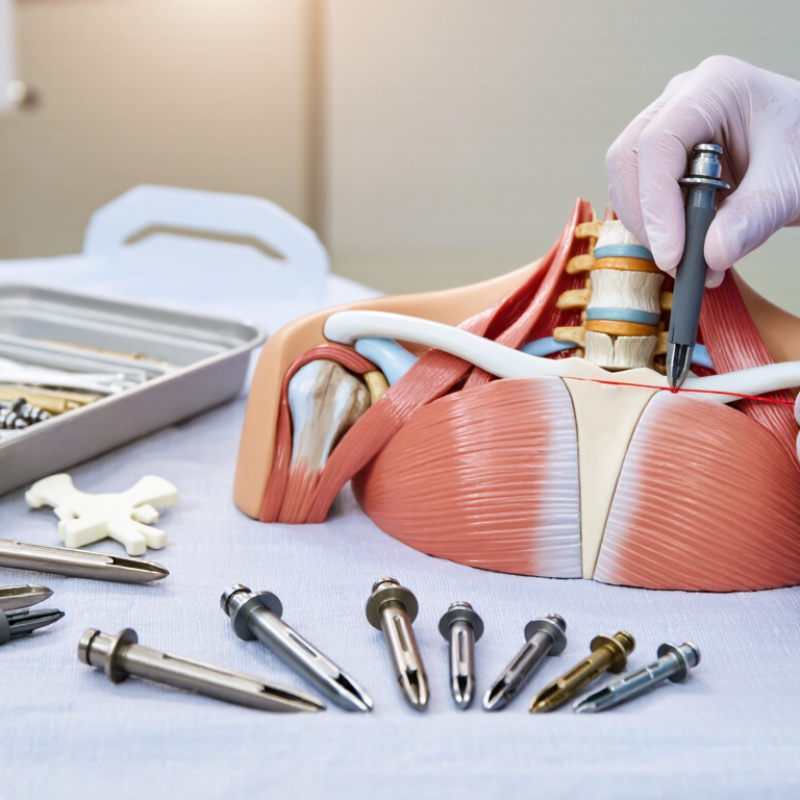
A suture anchor system is a small fixation device used to attach soft tissue to bone. It usually includes an anchor, high-strength suture, and insertion instrument. Surgeons place the anchor into prepared bone, then pass the suture through a tendon, ligament, or capsule. Tension restores the tissue’s intended position.
The design is more complex than its size suggests. Anchor material may be metallic, polymeric, or bioabsorbable. Each option affects visibility, handling, strength, and possible healing response. A 2024 industry market report estimates the global suture anchor market will grow at roughly 5–6% annually through 2030. Forecasts vary, however, because analysts define the market differently.
Clinical selection should begin with the repair, not the catalog. Consider bone quality, tissue thickness, anchor diameter, insertion angle, and required load. The suture must pass smoothly without damaging delicate tissue. The inserter should provide controlled depth and reliable deployment. Small details matter.
In practice, surgeons also evaluate pullout resistance, knot security, and imaging compatibility. Laboratory strength is useful, but it does not reproduce every operating-room condition. That is an important limitation. Peer-reviewed orthopedic reviews consistently emphasize matching fixation design to anatomy, loading, and surgical technique. A system can appear excellent on paper yet feel awkward during insertion. Careful training, documented outcomes, and transparent technical data remain essential.
Choosing the best suture anchor system starts with the operation, not the catalog.
The surgeon should assess tear pattern, tissue quality, bone density, and expected loading. A small, mobile tendon may need a different fixation strategy than a retracted tear with poor tissue. Fit matters. Anchor diameter, insertion depth, and eyelet position must suit the available bone.
Clinical evidence should guide the decision. A 2023 systematic review in Arthroscopy reported rotator cuff retear rates of roughly 15% to 30%, depending on tear size, patient factors, and follow-up duration. This does not prove that one anchor design prevents failure.
It shows how biology and repair technique remain central. The AAOS Clinical Practice Guideline for rotator cuff injuries also emphasizes patient-specific assessment and shared decision-making, rather than a universal implant choice. Think clinically.
Surgical access adds another requirement. In a narrow portal, the system should allow controlled deployment, reliable suture management, and clear confirmation of seating. For fragile bone, pullout resistance and insertion torque deserve careful attention.
A 2024 market analysis from Grand View Research projected continued growth in the global orthopedic biomaterials market, but market growth is not clinical proof. That distinction matters. The FDA’s MAUDE database can reveal device-related reports, yet passive reporting may miss events and cannot establish incidence.
Surgeons should combine published outcomes, regulatory information, hands-on experience, and the patient’s anatomy before selecting an anchor. Do not guess.
Choosing a suture anchor system starts with material behavior, not product appearance. Titanium offers high strength and clear radiographic visibility, but it can create imaging artifacts and may irritate surrounding tissue. PEEK is radiolucent and maintains structural stability, helping surgeons assess healing on follow-up scans. Its weakness is subtle: insertion damage may be harder to detect immediately.
Bioabsorbable polymers gradually break down. Their performance depends on hydrolysis, local biology, and the patient’s healing rate. A 2023 systematic review in Arthroscopy found no consistent clinical superiority between absorbable and nonabsorbable anchors across comparative studies. That finding deserves attention.
“Absorbable” does not automatically mean safer.
Design changes the fixation experience. Knotted anchors allow tension adjustment, while knotless systems reduce bulk and simplify final seating. All-suture anchors use smaller drill holes, which may preserve bone, but they require careful deployment and adequate cortical support.
A 2024 market analysis by Grand View Research projected steady growth in the global suture anchor market through 2030, reflecting wider use in sports medicine and soft-tissue repair. Market growth is not proof of better outcomes. Surgeons should compare pullout strength, insertion torque, imaging needs, bone quality, and peer-reviewed complication data before choosing. Small details matter.
Choosing a suture anchor system begins with the expected load, not the catalog label. Published biomechanical studies report initial failure loads ranging roughly from 100 to 300 N, depending on bone quality, anchor design, and testing method. These figures are not interchangeable. ASTM F543 testing also reminds clinicians that laboratory pullout strength may not represent cyclic loading in a shoulder or hip.
Strength should be matched to tissue and fixation purpose. A larger anchor may improve purchase in soft bone, yet it can remove more bone and complicate revision. The 2022 AAOS Clinical Practice Guideline emphasizes that repair decisions should reflect tear pattern, tissue quality, and patient factors, rather than one device feature. Measure insertion depth carefully. Check whether the anchor remains fully seated. Small details matter.
Placement deserves equal attention. Keep the anchor away from cartilage, fracture lines, and vulnerable neurovascular structures. Use the planned angle, then confirm the trajectory under direct visualization. Spacing also matters; crowded anchors can weaken the repair construct. A 2023 review of biomechanical literature found that cyclic loading often exposes differences missed by single-pull tests. That finding should change how systems are compared. This is where judgment remains imperfect. Bone density, surgeon technique, and rehabilitation can alter outcomes beyond the test bench. A technically strong anchor cannot rescue poor placement.
Choosing the best suture anchor system requires more than comparing sizes or stated strength. Safety must guide every decision.
Safety begins with reviewing the patient’s anatomy, bone quality, surgical site, and healing risks. Experienced teams confirm the anchor’s material and the patient’s allergy history. They also check imaging compatibility, especially when future magnetic resonance scans may be needed. Do not assume all materials behave alike. This is easy to miss.
Compatibility depends on the repair method and the tissue being secured. Match the anchor diameter, insertion depth, and suture configuration to the available bone and surgical space. Soft or fragile bone may not provide enough purchase for a standard design. The correct choice can change during surgery. Suture thickness, knot security, and abrasion resistance also deserve close attention. A small mismatch can weaken the repair or damage delicate tissue.
Before use, verify packaging integrity, sterility, expiration dates, and the instructions for the specific system. Confirm that the inserter fits firmly and that the deployment steps are clear to the operating team. During insertion, unusual resistance, poor seating, or unexpected cracking should prompt reassessment. Do not force it. Record the selected size, material, and lot information for reliable follow-up. A checklist helps, but it cannot replace clinical judgment. That limitation matters. The safest decision considers both documented specifications and the patient’s real anatomy.
The chart groups the essential checks required before selecting and using a suture anchor system. The values represent the number of verification items in each category, including patient factors, anatomy, implant compatibility, instrumentation, and postoperative planning. Always confirm the device-specific instructions for use, contraindications, MRI conditions, sterilization requirements, and surgical technique before use.
Start with the operation, not the catalog. Assess tear pattern, tissue quality, bone density, and expected loading. A small mobile tendon may need a different fixation strategy. A retracted tear with poor tissue requires closer planning. Fit matters.
Anchor diameter, insertion depth, and eyelet position must match the available bone. Limited bone space may require a smaller or differently positioned anchor. The repair should sit securely without damaging nearby tissue. Small mismatches can matter.
Current evidence does not prove that one design prevents failure. Retear rates vary with tear size, patient factors, healing biology, and follow-up time. Surgical technique remains important. The ideal choice is not always obvious.
The system should permit controlled deployment and reliable suture management. The team should clearly confirm seating. Crowded access can make handling difficult. Do not guess.
Fragile bone may provide limited purchase for a standard design. Review pullout resistance and insertion torque carefully. Watch for unusual resistance or cracking during insertion. Do not force it.
Review anatomy, bone quality, surgical-site conditions, healing risks, and allergy history. Confirm the material and future imaging compatibility. Do not assume all materials behave alike. This is easy to miss.
Inspect packaging integrity, sterility, and expiration dates. Confirm the inserter fits firmly and the deployment steps are understood. Keep the instructions available for the specific system. Even careful teams can miss a detail.
Record the selected size, material, and lot information. Document unusual resistance, poor seating, or other concerns. A checklist helps, but it cannot replace judgment. The decision may change during surgery.
Choosing the best Suture Anchor System requires a clear understanding of its role in securing soft tissue to bone during orthopedic and related surgical procedures. Selection should begin with the surgical objective, tissue quality, anatomical location, fixation method, and the surgeon’s preferred technique. Anchors may differ in material, absorption profile, thread configuration, eyelet structure, and deployment design, so each option should be evaluated according to the demands of the repair rather than by general performance claims.
Strength, size, and placement are also essential considerations. The anchor should provide sufficient initial fixation while remaining appropriate for the available bone and surrounding anatomy. Its dimensions, insertion angle, and spacing must support stable repair without creating unnecessary tissue or bone damage. Before use, clinicians should verify compatibility with the intended sutures, instruments, imaging requirements, and sterilization procedures. Careful assessment of handling characteristics, safety information, and applicable clinical guidance can help ensure that the selected system supports reliable surgical performance and patient-specific needs.